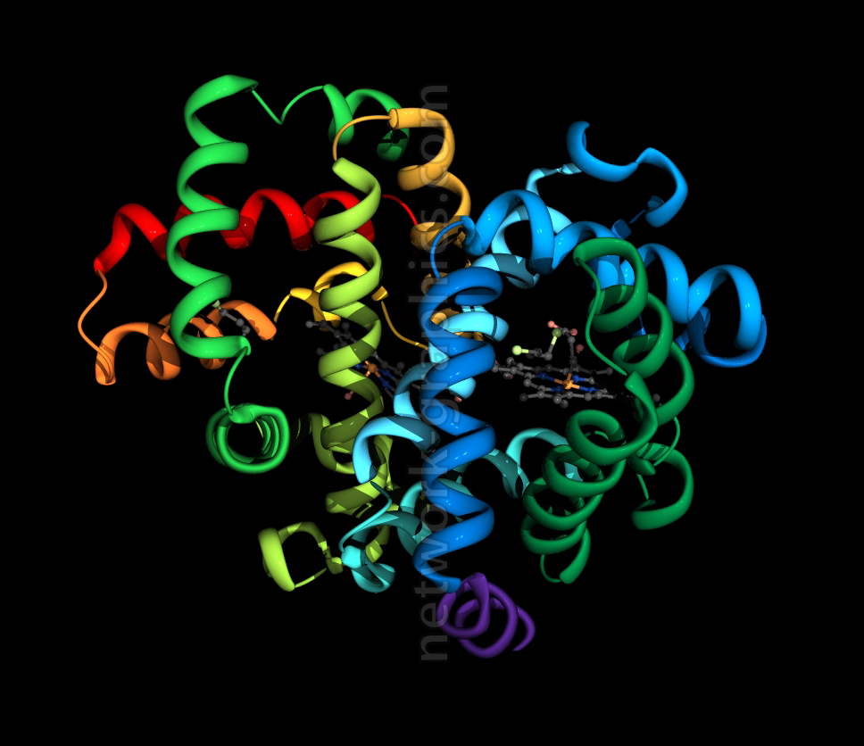
Hemoglobin Protein Structure with Bound Heme Group.

This 3D model depicts the tertiary structure of hemoglobin, a crucial oxygen-transport protein found in red blood cells. The image shows the alpha helices and beta sheets that fold to form the complex structure of hemoglobin, colored in a rainbow spectrum to indicate different chains or regions. At the core of the structure, the heme group (shown in dark gray) is clearly visible, highlighting its role in oxygen binding through the iron ion (Fe²⁺) at its center.
This image is ideal for biochemistry textbooks and molecular biology educational materials that focus on protein structure, enzyme function, and oxygen transport mechanisms in the human body.
We can provide sample images or create custom illustrations tailored to your projects. If you are looking for an illustration of this type, or from another subject area, you can contact us to discuss your needs.
Network Graphics / Division of Abramson & Wickham Graphics Inc.
All rights reserved.

