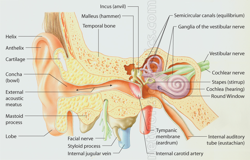
Cross-section of the Human Ear.

This color illustration provides a comprehensive cross-sectional view of the human ear, depicting its intricate anatomical structures. It includes clearly labeled parts of the outer, middle, and inner ear. The outer ear is shown with the helix, antihelix, concha (bowl), and external acoustic meatus leading into the ear canal. The middle ear highlights the malleus (hammer), incus (anvil), and the tympanic membrane (eardrum), which transmit sound vibrations to the inner ear. Also illustrated is the stapes (stirrup) connecting to the cochlea, responsible for hearing, and the vestibular nerve, which plays a role in balance and equilibrium.
Additional key features include the semicircular canals and ganglia of the vestibular nerve, as well as the facial nerve, internal jugular vein, and internal carotid artery running through the region. This detailed medical diagram serves as an educational resource for understanding ear anatomy, making it useful for textbooks, scientific publications, and e-learning.
We can provide sample images or create custom illustrations tailored to your projects. If you are looking for an illustration of this type, or from another subject area, you can contact us to discuss your needs.
Network Graphics / Division of Abramson & Wickham Graphics Inc.
All rights reserved.

