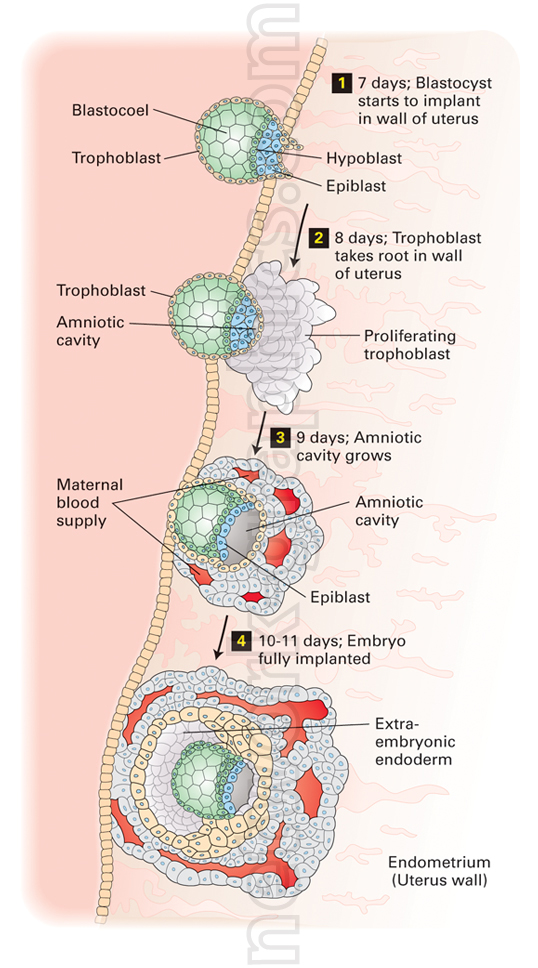
Blastocyst implantation in the
uterus wall.

This detailed illustration shows the process of blastocyst implantation into the uterus wall (endometrium), capturing the stages of early embryonic development. The blastocyst initially attaches to the uterus wall, with key components such as the blastocoel, trophoblast, epiblast, and hypoblast visible. Over time, the trophoblast expands and penetrates the uterine tissue, anchoring the developing embryo. As the implantation progresses, the amniotic cavity forms, providing a protective environment for the embryo, while the proliferating trophoblast interacts with the maternal blood supply to support growth.
In the final stages of implantation, the embryo becomes fully embedded within the endometrium. The extra-embryonic endoderm and amniotic structures are fully established, ensuring the embryo is securely implanted and ready for continued development. This high-quality and informative illustration provides a comprehensive view of the early stages of pregnancy and implantation.
This diagram is ideal for medical textbooks, biology journals, e-books, or educational articles covering topics related to embryology, human reproduction, and developmental biology.
We can provide sample images or create custom illustrations tailored to your projects. If you are looking for an illustration of this type, or from another subject area, you can contact us to discuss your needs.
Network Graphics / Division of Abramson & Wickham Graphics Inc.
All rights reserved.

